The retina and pupil are two essential components of the human eye, each playing a distinct role in the visual process. This article explores the anatomy, function, and significance of the retina and pupil, shedding light on their intricate roles in vision. Whether it’s capturing light or regulating the amount entering the eye, these structures work harmoniously to facilitate sight. Understanding their functions is crucial for comprehending. vision-related conditions and seeking appropriate care from a Retina specialist in Dubai.
What is Retina of the eye?
The Retina: Vision’s Canvas
Structure and Location
The retina is a thin, delicate layer of tissue located at the back of the eye, lining the inner surface of the eyeball. It is often likened to the film of a camera, as it captures incoming light and converts it into neural signals that are sent to the brain for interpretation. Despite its diminutive size, the retina is incredibly complex, containing multiple layers of specialized cells that work together to facilitate vision.
Can Retina Heal Without Surgery?
Layers and Components
The retina comprises several layers, each with specific functions crucial for visual processing. The outermost layer, the retinal pigment epithelium (RPE), provides support to the photoreceptor cells and helps maintain the retina’s health. The photoreceptor layer contains two types of cells: rods and cones. Rods are responsible for vision in low-light conditions, while cones enable color vision and sharp visual acuity. Bipolar cells and ganglion cells further process visual information before transmitting it to the brain via the optic nerve.
The Pupil: Gateway to Vision
Anatomy and Function
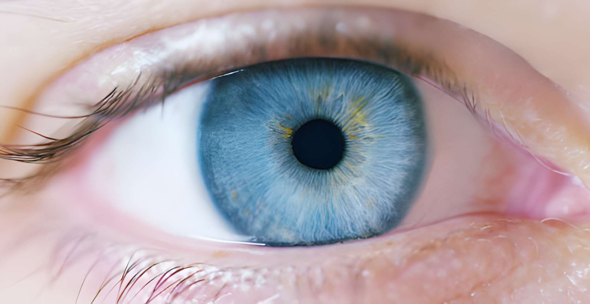
The pupil is the black circular opening at the center of the iris, the colored part of the eye. It appears black because light entering the eye is absorbed by the retina and not reflected back. The pupil’s primary function is to regulate the amount of light entering the eye, thereby controlling the intensity of the visual stimuli reaching the retina. This process, known as pupillary reflex, helps maintain optimal visual clarity under varying lighting conditions.
How do I know if my retina is damaged?
Dynamic Adjustments
The size of the pupil is not fixed but rather dynamically adjusts in response to changes in ambient light levels and visual demands. In bright conditions, the pupil constricts or becomes smaller to limit the amount of light entering the eye and prevent glare. Conversely, in dim lighting, the pupil dilates or enlarges to allow more light to enter, enhancing visual sensitivity. These automatic adjustments ensure that the retina receives an appropriate amount of light for optimal visual performance.
Visual Process: From Retina to Brain
Light Capture and Conversion
When light enters the eye through the pupil, it passes through the cornea and lens, which focus it onto the retina. The photoreceptor cells in the retina then capture the light and convert it into electrical signals through a process called phototransduction. Rods and cones respond differently to light stimuli, with rods being more sensitive to low-light conditions and cones being responsible for color vision and detail.
Can Retina Problems be Cured?
Neural Signaling
The electrical signals generated by the photoreceptor cells are transmitted to the bipolar cells, which further process the visual information before transmitting it to the ganglion cells. The ganglion cells then bundle together to form the optic nerve, which carries the visual signals from the retina to the brain. Once in the brain, these signals are interpreted and translated into meaningful visual perceptions, allowing us to see and interact with the world around us.
Common Retinal and Pupillary Conditions
Retinal Disorders
Various conditions can affect the structure and function of the retina, leading to vision problems or loss. Age-related macular degeneration (AMD), diabetic retinopathy, retinal detachment, and retinitis pigmentosa are examples of retinal disorders that can cause significant visual impairment if left untreated. Consulting a retina specialist in Dubai is essential for accurate diagnosis and personalized treatment options tailored to each patient’s needs.
What Vitamin Deficiency is Retina?
Pupillary Abnormalities
Pupillary abnormalities can manifest as irregularities in the size, shape, or responsiveness of the pupil. Conditions such as anisocoria (unequal pupil size), Horner’s syndrome (pupil constriction), and Adie’s tonic pupil (dilated and sluggish pupil) can indicate underlying neurological or ocular issues. Prompt evaluation by an eye specialist is necessary to determine the cause of pupillary abnormalities and initiate appropriate management.
Diagnostic and Treatment Approaches
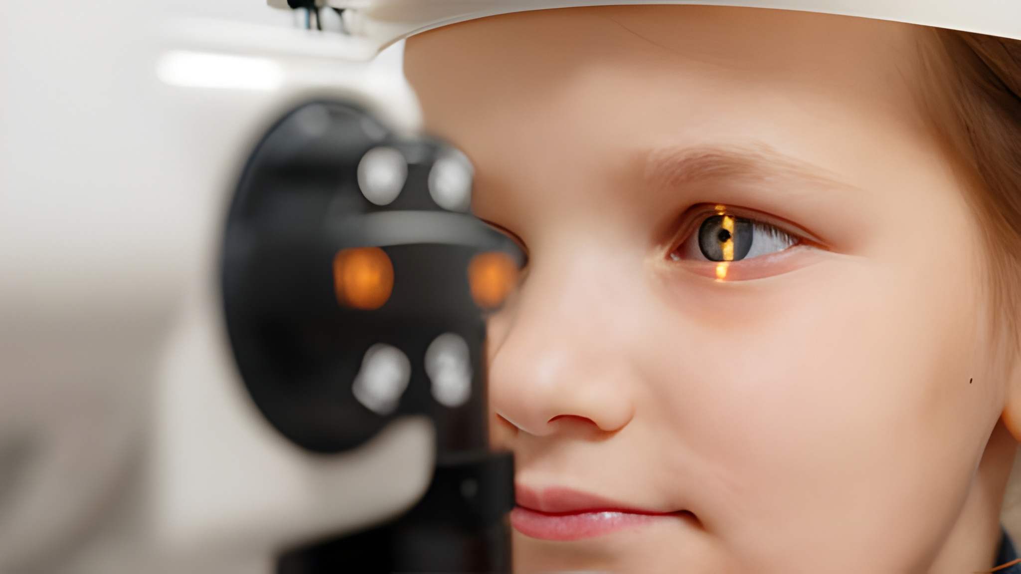
Retinal Imaging
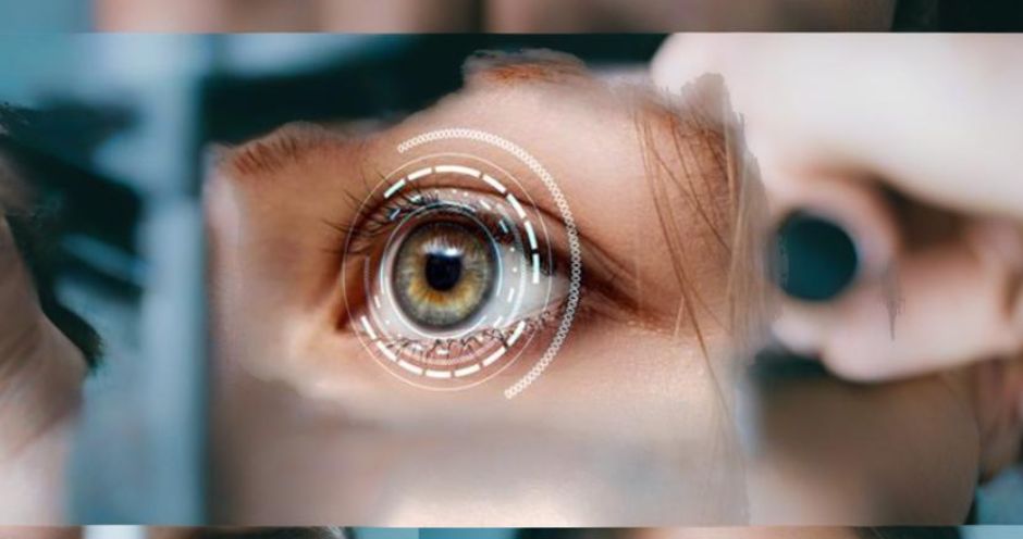
Retinal imaging techniques, such as optical coherence tomography (OCT) and fundus photography, allow for detailed visualization of the retina’s structure and integrity. These non-invasive imaging modalities provide valuable information for diagnosing retinal conditions, monitoring disease progression, and assessing treatment efficacy. Early detection through retinal imaging can facilitate timely intervention and preservation of vision.
What is the Cost of Retina Surgery in UAE
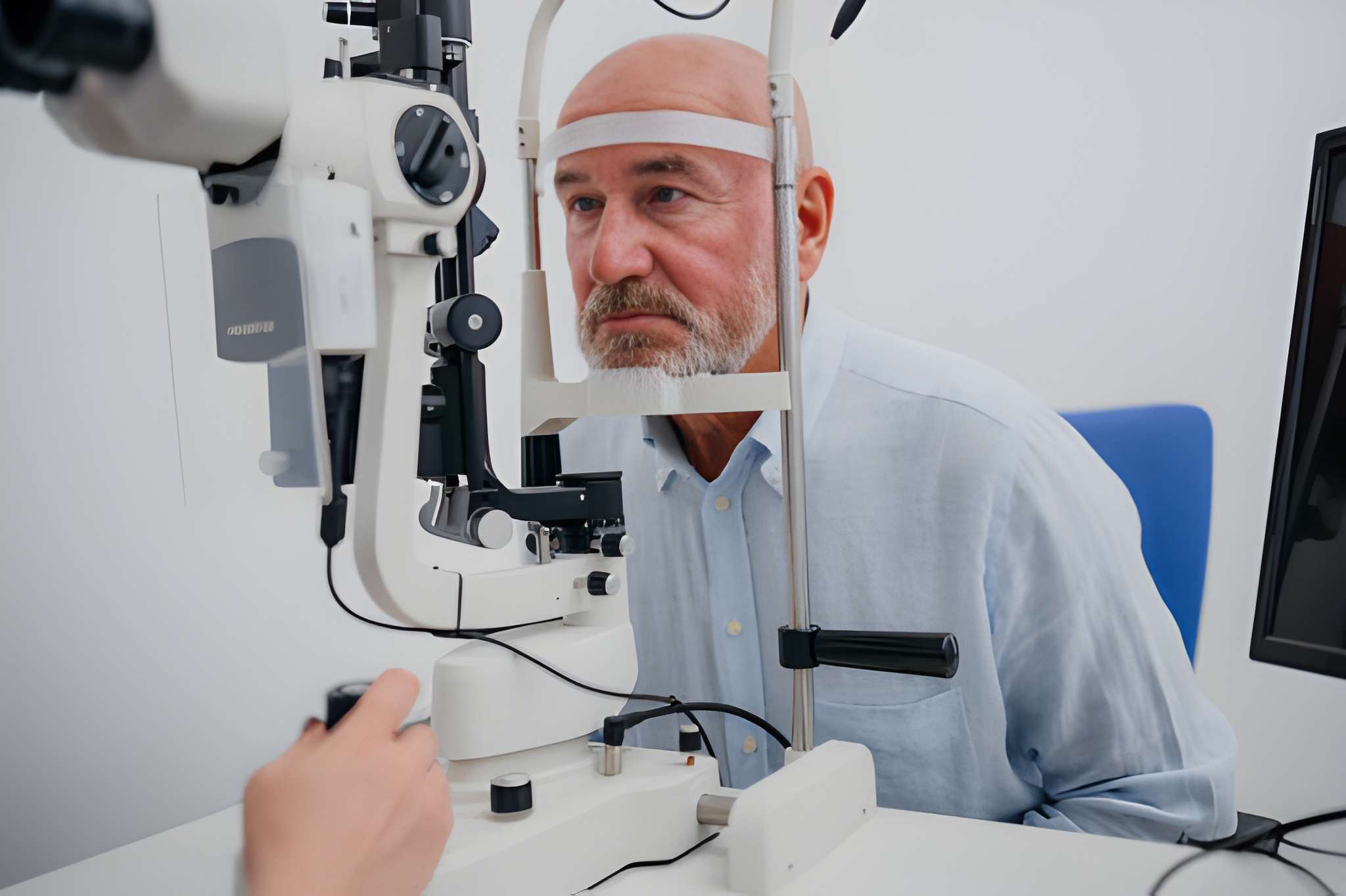
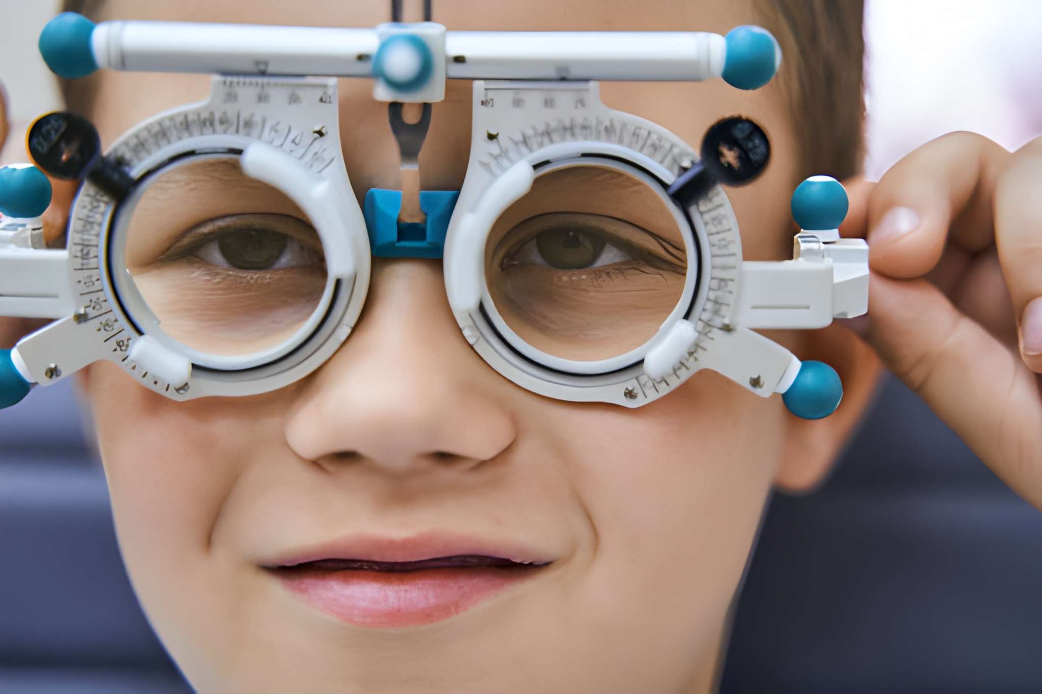
Pupillary Assessment
Pupillary assessment involves evaluating the size, symmetry, and reactivity of the pupils to light and accommodation. This assessment can help identify pupillary abnormalities and guide further diagnostic investigations. Pupillary responses are typically tested using a penlight or specialized pupillometer. Abnormal findings may indicate underlying neurological conditions, ocular trauma, or systemic disorders requiring further evaluation and management.
Seeking Specialized Care in Dubai
Importance of Eye Specialist
For individuals experiencing retinal or pupillary issues, seeking care from an eye specialist in Dubai is crucial. These highly trained professionals possess expertise in diagnosing and treating a wide range of ocular conditions, including those affecting the retina and pupil. From comprehensive eye examinations to advanced imaging techniques and specialized treatments, eye specialists offer personalized care to address each patient’s unique needs.
Is Retina Surgery Painful?
Retina Specialist in Dubai
A retina specialist in Dubai specializes in the diagnosis and management of retinal disorders, utilizing advanced diagnostic tools and treatment modalities to preserve and restore vision. Whether it’s AMD, diabetic retinopathy, or retinal detachment, a retina specialist can provide comprehensive care tailored to each patient’s condition and visual goals. Early intervention by a retina specialist can significantly impact treatment outcomes and visual prognosis.
Consult Dr. Qasim Qasem, Leading Eye Specialist in Dubai
For expert evaluation and personalized care for retinal and pupillary conditions, trust Dr. Qasim Qasem, a renowned eye specialist in Dubai. With extensive experience and a commitment to excellence, Dr. Qasim Qasem provides comprehensive eye care services, including diagnosis, treatment, and management of retinal and pupillary disorders. Utilizing state-of-the-art technology and a patient-centered approach, Dr. Qasim Qasem ensures that each patient receives the highest quality of care and achieves optimal visual outcomes. Contact Dr. Qasim Qasem today to schedule a consultation and take the first step toward better eye health and vision.
What are the symptoms of a weak retina?