The retina, a delicate and complex layer of tissue lining the back of the eye, plays a pivotal role in the process of vision. It serves as the gateway through which light enters the eye and is transformed into neural signals that the brain interprets as images. Understanding the structure and function of the retina is crucial for comprehending the mechanisms underlying vision and for diagnosing and treating various eye conditions. Without a properly functioning retina, our ability to perceive the world around us would be severely compromised. This article will delve into the intricate anatomy of the retina, its fundamental role in vision, the different types of cells comprising it, and the significance of maintaining retinal health. For those seeking exceptional eye care, look no further than Dr.Qasim Qasem, one of the best eye specialist in Dubai, renowned for their expertise and commitment to patient well-being.
Anatomy of the Retina
The retina is a multi-layered structure consisting of specialized cells that work together to process visual information. At the front of the retina lies the photoreceptor layer, which contains the cells responsible for detecting light: rods and cones. Rods are sensitive to dim light and are crucial for night vision, while cones are responsible for color vision and function best in bright light. Behind the photoreceptor layer is the bipolar cell layer, which receives signals from the photoreceptors and relays them to the ganglion cell layer. Ganglion cells collect and integrate visual information before transmitting it to the brain via the optic nerve. Additionally, the retina contains horizontal and amacrine cells, which help in processing visual signals within the retina itself.
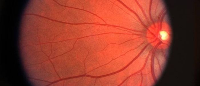
The blood supply to the retina is provided by two main sources: the central retinal artery and the choroid. The central retinal artery branches off from the ophthalmic artery and supplies oxygen and nutrients to the inner layers of the retina. The choroid, located behind the retina, supplies blood to the outer layers. This dual blood supply is essential for maintaining the metabolic needs of the retina and ensuring its proper function.
Understanding the intricate layers of the retina provides insight into how visual information is processed and transmitted within the eye.
Book Consultation With: Retina Specialist in Dubai
Function of the Retina
The retina serves as the initial site of visual processing, where light entering the eye is converted into electrical signals that can be interpreted by the brain. This process begins when light passes through the cornea and lens, focusing onto the retina. The photoreceptor cells in the retina then capture this light and convert it into electrical impulses. Rods and cones contain photopigments that undergo chemical changes when exposed to light, initiating a cascade of events that ultimately leads to the generation of neural signals.
These neural signals are then processed by the various layers of the retina, where they are refined and organized before being transmitted to the brain via the optic nerve. The brain interprets these signals, allowing us to perceive the visual world around us. This intricate process enables us to distinguish shapes, colors, and movement, forming the basis of our visual experience.
Understanding the role of the retina in vision provides valuable insights into how we perceive the world and the mechanisms underlying visual perception.
What are the Symptoms of a Week Retina
Photoreceptors: Rods and Cones
Photoreceptor cells, namely rods and cones, are specialized cells located in the outermost layer of the retina responsible for detecting light stimuli. Rods are highly sensitive to light and are primarily responsible for vision in low-light conditions, such as night vision. They contain a photopigment called rhodopsin, which undergoes a chemical change when exposed to light, initiating the process of signal transduction.
Cones, on the other hand, are responsible for color vision and function best in bright light. There are three types of cones, each sensitive to different wavelengths of light corresponding to the colors red, green, and blue. This allows us to perceive a wide spectrum of colors and differentiate between them. Unlike rods, cones contain photopigments such as photopsin, which are responsible for color vision.
Rods are more abundant in the peripheral regions of the retina, while cones are concentrated in the central region known as the fovea. This distribution reflects the different visual needs of the human eye, with the fovea specialized for high-acuity vision and color discrimination.
Understanding the differences between rods and cones and their distribution within the retina provides insights into how the visual system adapts to different lighting conditions and perceives color.
What Vitamin Deficiency is Retina?
Retinal Pigment Epithelium (RPE)
The retinal pigment epithelium (RPE) is a layer of pigmented cells located between the photoreceptor layer and the choroid, a vascular layer supplying blood to the retina. Despite its thinness, the RPE plays a crucial role in supporting the function and health of the retina.
One of the primary functions of the RPE is to provide nutritional support to the photoreceptor cells. It transports essential nutrients, such as glucose and ions, from the blood vessels in the choroid to the photoreceptor cells, ensuring their metabolic needs are met. Additionally, the RPE is involved in the removal of waste products generated by photoreceptor cells during the process of vision. It phagocytoses or engulfs the shed outer segments of photoreceptor cells, preventing the buildup of debris within the retina.
Moreover, the RPE is integral to the visual cycle, a series of biochemical reactions that occur in the photoreceptor cells during the process of vision. It plays a key role in regenerating the visual pigment molecules required for photoreceptor function. This process involves the recycling of retinoids, a type of vitamin A derivative, which are essential for the synthesis of visual pigments in photoreceptor cells.
Furthermore, the RPE contributes to the maintenance of the blood-retina barrier, a selective barrier that regulates the passage of molecules between the blood vessels and the retina. This barrier helps protect the delicate neural tissue of the retina from harmful substances circulating in the blood, maintaining the homeostasis of the retinal microenvironment.
Overall, the RPE plays a vital role in supporting the function and health of the retina, ensuring its proper functioning and integrity.
Can Retina Problems be Cured?
Neural Circuitry of the Retina
The retina contains a complex network of neurons that process visual information before it is transmitted to the brain. This neural circuitry allows for the integration and refinement of visual signals, enhancing visual perception and sensitivity.
The primary pathway through which visual information travels within the retina is known as the vertical pathway. This pathway consists of three main cell types: photoreceptors, bipolar cells, and ganglion cells. Photoreceptors, namely rods and cones, capture light stimuli and convert them into electrical signals. These signals are then transmitted to bipolar cells, which serve as intermediaries, relaying the signals from photoreceptors to ganglion cells. Ganglion cells are the output neurons of the retina, sending visual information to the brain via the optic nerve.
In addition to the vertical pathway, the retina contains a horizontal pathway that allows for lateral communication between adjacent neurons. Horizontal cells receive input from multiple photoreceptors and provide inhibitory feedback to regulate the activity of surrounding cells. This lateral inhibition enhances contrast and sharpens spatial information in visual processing.
Amacrine cells, another type of interneuron found in the retina, play a crucial role in modulating the transmission of signals between bipolar and ganglion cells. They are involved in various functions, such as temporal filtering, spatial integration, and direction selectivity, contributing to the refinement of visual information.
The intricate neural circuitry of the retina allows for the processing and integration of visual signals, optimizing visual perception and sensitivity.
Can Retina Heal Without Surgery?
Retinal Disorders
The delicate structure and intricate function of the retina make it susceptible to various disorders and diseases that can impair vision. These conditions often arise from abnormalities in the retina’s structure, function, or blood supply, leading to visual disturbances and, in some cases, vision loss. Understanding common retinal disorders is crucial for early detection, diagnosis, and management to preserve vision.
Age-related Macular Degeneration (AMD): Age-related macular degeneration is a leading cause of vision loss in older adults. It affects the macula, the central part of the retina responsible for sharp, central vision. AMD can manifest as either dry AMD, characterized by the gradual breakdown of retinal cells and the accumulation of drusen (tiny deposits) beneath the retina, or wet AMD, marked by the growth of abnormal blood vessels beneath the macula. These blood vessels can leak fluid and blood, causing scarring and irreversible damage to the macula.
Diabetic Retinopathy: Diabetic retinopathy is a complication of diabetes that affects the blood vessels in the retina. High levels of blood sugar can damage the small blood vessels, causing them to leak fluid or bleed into the retina. In the early stages, diabetic retinopathy may not cause noticeable symptoms, but as the condition progresses, it can lead to vision loss or even blindness. Diabetic retinopathy can manifest as non-proliferative diabetic retinopathy (NPDR) or proliferative diabetic retinopathy (PDR), depending on the severity of the damage to the blood vessels.
Read More: Is Retina Surgery Painful?
Retinal Detachment: Retinal detachment occurs when the retina separates from the underlying tissue, disrupting its blood supply and causing vision loss. This condition often results from a tear or hole in the retina, allowing fluid to accumulate between the retina and the underlying layers. Retinal detachment is a medical emergency that requires prompt treatment to prevent permanent vision loss. Symptoms may include sudden flashes of light, floaters in the field of vision, and a curtain-like shadow or loss of vision in one eye.
Retinitis Pigmentosa: Retinitis pigmentosa is a group of inherited retinal disorders characterized by progressive degeneration of the photoreceptor cells in the retina. This condition typically begins with night blindness and peripheral vision loss, eventually leading to tunnel vision or complete blindness in severe cases. Retinitis pigmentosa can result from mutations in various genes involved in the structure and function of photoreceptor cells, affecting their ability to respond to light stimuli.
These are just a few examples of the many retinal disorders that can affect vision. Early detection, diagnosis, and treatment are essential for managing these conditions and preserving visual function.
Advances in Retinal Research
Advancements in retinal research have led to significant insights into the underlying mechanisms of retinal function and dysfunction, as well as novel approaches for diagnosing and treating retinal disorders. Researchers and clinicians continue to explore innovative techniques and therapies to improve outcomes for patients with retinal diseases.
Gene Therapy: Gene therapy holds promise for treating inherited retinal disorders by delivering functional genes to replace or supplement defective ones. Recent clinical trials have shown encouraging results in restoring vision in patients with conditions such as Leber congenital amaurosis and choroideremia. Gene editing technologies, such as CRISPR-Cas9, offer potential avenues for precise targeting and correction of genetic mutations associated with retinal diseases.
Stem Cell Therapy: Stem cell therapy aims to regenerate damaged retinal tissue and restore visual function by transplanting stem cells into the retina. Researchers are investigating various sources of stem cells, including embryonic stem cells, induced pluripotent stem cells, and retinal progenitor cells, for their potential to differentiate into retinal cells and integrate into the existing neural circuitry.
Retinal Prostheses: Retinal prostheses, also known as retinal implants or bionic eyes, are devices designed to restore vision in individuals with severe vision loss or blindness. These implants bypass damaged retinal cells and directly stimulate the remaining functional cells or the visual cortex of the brain. While current retinal prostheses have limitations in terms of resolution and field of view, ongoing research aims to enhance their efficacy and accessibility for a wider range of patients.
What is the retina and pupil?
Optogenetics: Optogenetics is a cutting-edge technique that involves genetically modifying retinal cells to respond to light of specific wavelengths, enabling light-driven activation of neural circuits. This approach holds the potential for restoring vision in conditions where photoreceptor cells are degenerated or non-functional, such as retinitis pigmentosa. By introducing light-sensitive proteins into remaining retinal cells or neurons in the visual pathway, optogenetics offers a targeted and customizable strategy for vision restoration.
Artificial Intelligence (AI): Artificial intelligence and machine learning algorithms are increasingly being employed to analyze retinal images and detect early signs of retinal diseases. AI-driven diagnostic tools can assist clinicians in identifying subtle abnormalities in retinal morphology or vasculature, facilitating earlier intervention and treatment. Moreover, AI algorithms can aid in predicting disease progression and optimizing treatment strategies based on individual patient characteristics.
These advancements represent just a fraction of the ongoing research efforts aimed at understanding and treating retinal disorders. Continued collaboration between scientists, clinicians, engineers, and patients is essential for translating these innovations into clinically meaningful therapies that improve the lives of individuals affected by retinal diseases.
Importance of Retinal Health
Maintaining retinal health is crucial for preserving clear, sharp vision and overall visual function. Several factors contribute to retinal health, including lifestyle choices, regular eye examinations, and early detection of retinal disorders.
Lifestyle Factors: Adopting a healthy lifestyle can promote retinal health and reduce the risk of developing retinal diseases. This includes maintaining a balanced diet rich in antioxidants, vitamins, and omega-3 fatty acids, which support retinal function and protect against oxidative stress. Regular exercise and weight management can also help prevent conditions such as diabetes and hypertension, which are risk factors for diabetic retinopathy and other vascular-related retinal diseases.
Eye Examinations: Regular comprehensive eye examinations are essential for detecting early signs of retinal diseases and monitoring changes in vision over time. Eye care professionals can perform various tests to evaluate retinal health, including visual acuity testing, intraocular pressure measurement, and dilated fundus examination. Additional imaging modalities, such as optical coherence tomography (OCT) and fundus photography, provide detailed views of the retina and aid in diagnosing retinal disorders at their earliest stages.
Early Detection: Early detection of retinal disorders allows for timely intervention and treatment, minimizing the risk of irreversible vision loss. Symptoms such as blurry vision, floaters, flashes of light, or changes in peripheral vision should prompt immediate evaluation by an eye care professional. Routine screenings for conditions such as diabetic retinopathy are especially important for individuals at higher risk, such as those with diabetes or a family history of the disease.
By prioritizing retinal health and taking proactive measures to protect vision, individuals can maintain optimal visual function and enjoy a high quality of life.
Final Thoughts
The retina, an extraordinary structure nestled within the eye, serves as the cornerstone of vision. Its intricate architecture and multifaceted functionality empower us to navigate and interpret the world with remarkable clarity and precision. Delving into the intricacies of the retina, encompassing its diverse layers, cellular constituents, and intricate neural pathways, unveils profound insights into the mechanisms orchestrating visual perception and the underlying pathology of retinal ailments. From capturing minute details to discerning hues and motion, the retina orchestrates an intricate symphony of sensory experiences that shape our perception of reality.
Consult with Ophthalmologist Dr. Qasim Qasem Today!
Dr. Qasim Qasem, a highly skilled and experienced ophthalmologist, is dedicated to providing exceptional eye care services tailored to your needs. Whether you’re experiencing vision problems, eye discomfort, or simply seeking preventive care, Dr. Qasim Qasem is here to help. With expertise in diagnosing and treating a wide range of eye conditions, including cataracts, glaucoma, and retinal disorders, Dr. Qasim Qasem offers personalized treatment plans to optimize your visual health.
Book Appointment With Dr. Qasim Today


